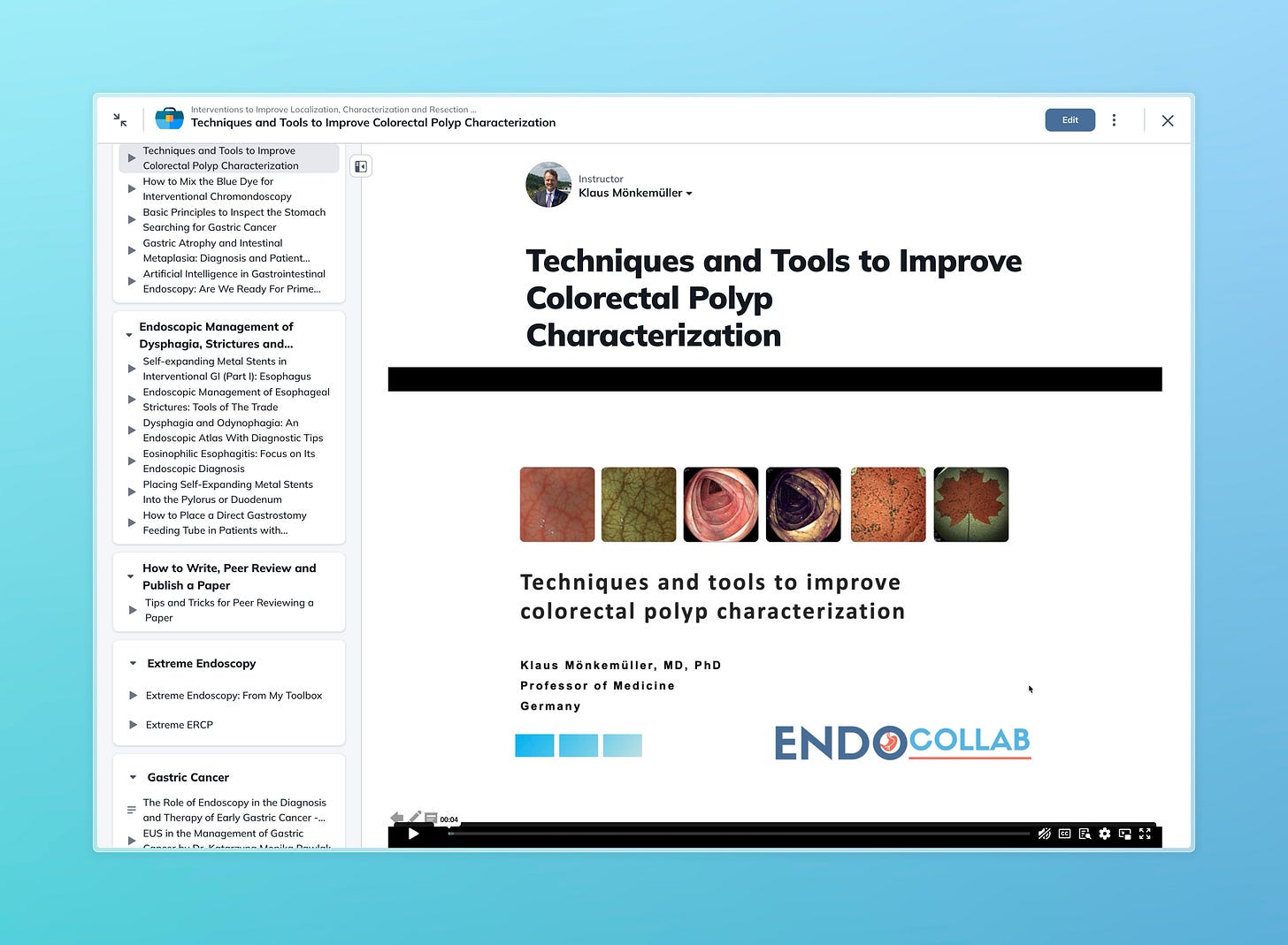
Techniques and Tools to Improve Colorectal Polyp Characterization
EndoCollab Weekly Digest: March 1-15, 2025
Join us as we dive into essential techniques and cutting-edge tools—from the Paris and Kudo classifications to virtual chromoendoscopy and AI-assisted diagnostics. Whether you're refining your skills or exploring the latest innovations, this lecture offers practical insights to elevate your endoscopic practice. Watch now and train your eye to spot what matters!"
What’s Covered in the Video:
Fundamentals of Polyp Inspection: Learn the basics of evaluating polyp shape, surface (pit pattern), and capillary network for accurate characterization.
Paris Classification: Understand polyp morphology with examples like pedunculated (1P), sessile (1S), flat (2A/2B), and excavated (3C) lesions, including laterally spreading tumors (LSTs).
Kudo Pit Pattern Classification: Explore pit patterns (Type 1 to 5) and their correlation with normal mucosa, hyperplastic lesions, adenomas, and carcinomas.
Sano Vascular Classification: Assess vascular patterns to gauge disease severity, from non-neoplastic (Type 1) to carcinoma (Type 3).
NICE Classification: Master the Narrow Band Imaging International Colorectal Endoscopic Classification for quick, practical polyp assessment (Type 1: hyperplastic, Type 2: adenoma, Type 3: cancer).
Traditional Chromoendoscopy: Discover the use of contrast dyes (e.g., indigo carmine) and absorptive dyes (e.g., crystal violet) to enhance polyp detection and characterization.
Interventional Chromoendoscopy: See how submucosal dye injections improve resection outcomes, with real case examples from 15 years of practice.
Dye-Less Virtual Chromoendoscopy: Get insights into NBI, iScan, BLI, and LCI—modern tools for characterizing lesions without traditional dyes.
Detection vs. Characterization: Understand how advanced imaging boosts both polyp detection and diagnostic precision, backed by studies.
Emerging Technologies: Preview molecular imaging and AI-assisted diagnosis, including their potential to revolutionize endoscopy.
Practical Tips: Train your eye with expert advice on improving diagnostic accuracy using online resources and visual practice.
Case Examples: Real endoscopic images and resection techniques (e.g., EMR, ESD) to bridge theory and practice.
WhatsApp Weekly Digest:
Below is a summary of the key cases, learnings, and recommendations from the Endocollab WhatsApp group transcript spanning March 1 to March 15, 2025. This is tailored for busy group members who couldn’t follow the full conversation, highlighting the main cases, along with the key insights and advice shared.
Summary of Key Cases and Learnings (March 1–15, 2025)
Case 1: 85-Year-Old Man with Gastric Outlet Obstruction
Presentation: Elderly male with gastric outlet obstruction, hypokalemia, and UGIE showing D2 stenosis. Esophagus showed Los Angeles Grade D esophagitis.
Key Learning: Severe esophageal damage (Grade D) was noted alongside duodenal stenosis, suggesting a complex upper GI pathology.
Recommendations: No specific treatment recommendations were detailed, but the case underscores the need to assess both esophageal and duodenal findings in such presentations.
Case 2: 17-Year-Old Male with Rectal Lesion
Presentation: Lesion 12–13 cm above the rectum, with abdominal pain and occasional bloody stools.
Key Learning: Differential included solitary rectal ulcer (suggested with biopsy correlation) and possible colonic Crohn’s disease.
Recommendations: Awaiting histopathology; clinical history and biopsy are critical for diagnosis. Crohn’s was considered a possibility, emphasizing the need for thorough investigation.
Case 3: 19-Year-Old Male with Antral Mass
Presentation: Abdominal pain for 1 week; CT and OGD showed an antral mass. Repeat OGD/EUS after PPI showed a mass with mucous discharge, disrupted wall layers, and benign cytology.
Key Learning: Ectopic pancreas with possible malignant transformation was suspected, though biopsies were negative. Pancreatic rest and hydatid cyst were also considered.
Recommendations: No intervention if asymptomatic; further imaging (e.g., EUS) to clarify etiology. Follow-up was requested to monitor transformation post-PPI.
Case 4: 30-Year-Old Male with Recurrent Acute Pancreatitis
Presentation: Recurrent pancreatitis with biliary etiology (post-cholecystectomy); no other causes identified (e.g., alcohol, genetic, obstructive).
Key Learning: Biliary pancreatitis was the likely trigger; comprehensive exclusion of alternative causes was emphasized.
Recommendations: The team’s diagnostic approach was endorsed as thorough; no new treatment suggestions were added beyond prior management.
Case 5: 30-Year-Old Female with Pancreas Divisum and Pancreatic Stones
Presentation: Chronic pancreatitis with pancreas divisum, multiple pancreatic duct stones, and pain.
Key Learning: Stone size dictates management—small stones (<5 mm) can be removed via ERCP, while larger ones may require lithotripsy or surgery.
Recommendations: ERCP for stones <5 mm; extracorporeal shock wave lithotripsy (ESWL) or Puestow procedure for larger stones. UDCA was not recommended.
Case 6: 33-Year-Old Female with Pancreatic Calculi
Presentation: Calculi from pancreatic head to tail, recurrent epigastric pain.
Key Learning: Extensive calculi may require more aggressive intervention.
Recommendations: Surgery or ERCP with SpyGlass was suggested for stone removal, tailored to symptom severity and technical feasibility.
Case 7: 10-Year-Old with Duodenal Varices and EHPVO
Presentation: Portal hypertension from extrahepatic portal vein obstruction (EHPVO) with eradicated esophageal varices; non-bleeding duodenal varices noted.
Key Learning: Primary prophylaxis for non-bleeding varices lacks robust data in EHPVO; secondary prophylaxis aligns with cirrhosis guidelines.
Recommendations: Glue injection for bleeding varices; beta-blockers (e.g., carvedilol) and band ligation for secondary prophylaxis if bleeding history exists. Rex shunt was recommended for growth retardation in this pediatric case.
Case 8: Young Male with Suspected Pancreatic Rest
Presentation: Epigastric pain with endoscopic findings suggestive of pancreatic rest in the stomach.
Key Learning: Pancreatic rests are usually asymptomatic; malignancy is rare but suspected with atypical endoscopic features (e.g., irregular margins).
Recommendations: Leave asymptomatic lesions alone; EUS to confirm ductal structure if malignancy is a concern. Resection only if symptomatic or complicated.
Case 9: 30-Year-Old Male with Bile Reflux Gastritis
Presentation: Dyspepsia with bile in the stomach due to an anomalous left hepatic duct opening into the antrum.
Key Learning: Bile reflux caused reactive gastropathy, not chronic gastritis; pancreatic secretions may contribute to mixed gastritis.
Recommendations: UDCA and prokinetics resolved symptoms; mucosa healed after 2 months. Patient remained asymptomatic at 2-year follow-up, suggesting a functional component to pain.
General Insights
Diagnostic Tools: EUS and biopsy were frequently recommended to clarify ambiguous findings (e.g., pancreatic rest, antral masses).
Intervention Threshold: Asymptomatic lesions (e.g., ectopic pancreas) are best left alone, adhering to "primum non nocere" (first, do no harm).
Join our discussion in the EndoCollab WhatsApp Group. Join now!


