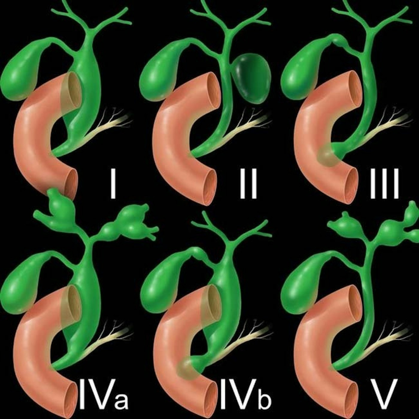
Todanic Classification of Choledocal Cysts
Understanding Choledochal Cysts: A Comprehensive Classification Guide to Biliary Tract Anomalies
Type I: Solitary fusiform or cystic dilation of extrahepatic bile duct
Type Ia: Cystic dilation of entire extrahepatic bile duct; associated with abnormal pancreaticobiliary junction (APBJ)
Type Ib: Focal dilation of extrahepatic bile duct (often distal); no association with APBJ Type
Ic: Fusiform dilation of entire extrahepatic bile duct; associated with APBJ. This is the most common type, constituting 50-85% of choledochal cysts. Much more common in females than males (3:1) and may present with pain, jaundice, or gallstone formation (due to bile stasis). You need to differentiate from distal obstruction of extrahepatic bile duct obstruction (e.g., stone or tumor). Mild dilation of right and left ducts may blur distinction with type IVa.
Type II: True diverticulum of supraduodenal extrahepartic bile duct, accounting for only 2% of cases
Type III: Dilation limited to intraduodenal segment of extrahepatic bile duct (choledochocele), with dilated segment of extrahepatic bile duct located within duodenal wall
Type IIIa: Cystic dilation of intraduodenal extrahepatic bile duct
Type IIIb: Diverticulum of intraduodenal extrahepatic bile duct. Constitutes 1-5% of cases. Cyst may be lined by either duodenal or biliary epithelium. Large choledochoceles may obstruct duodenum, present with jaundice, or cause pancreatitis.
Type IV: Presence of multiple biliary cysts, at least 1 of which must involve extrahepatic bile duct
IVa: Involvement of both intrahepatic and extrahepatic ducts; second most common overall, comprising 40% of cases diagnosed in adults IVb: Multiple extrahepatic cysts with no intrahepatic cysts. Constitutes 15-35% of cases.
Type V: Single or multiple intrahepatic biliary cysts, with presence of multiple intrahepatic cysts known as Caroli disease No involvement of extrahepatic duct.
Reference: https://radiologykey.com/choledochal-cyst/


