

WhatsApp Chat Summary: September 113, 2025 → September 30, 2025
From advanced imaging techniques and polyp management to portal hypertension, radiation complications, genetic syndromes, and hemostasis strategies.
Welcome back to the EndoCollab Newsletter! This edition summarizes the key discussions and teaching points from our WhatsApp group over the latter half of September. We’ve explored a range of challenging cases and shared valuable insights, which this newsletter aims to consolidate for our collective learning and reference.
If you’d like to participate in our WhatsApp discussions, you can do so by becoming a lifetime member of EndoCollab here.
Case Chapters
Case 1: The Nuances of Rectal Polyp Assessment
Case 2: A Suspected Granulomatous Polyp
Case 3: Extensive Colonic Varices in a Young Woman
Case 4: Strong Caution Against APC After Brachytherapy
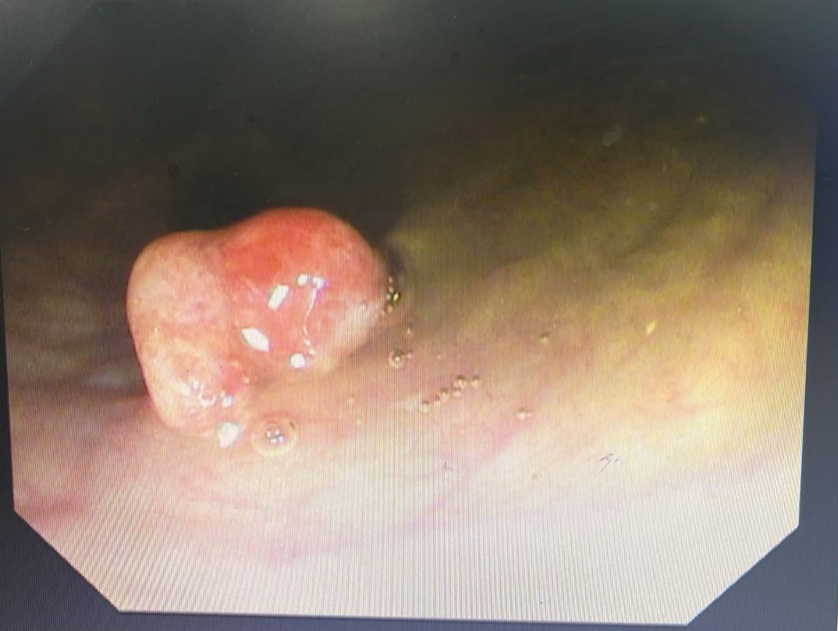
Case 5: The Dilemma of the Gastric Hyperplastic Polyp
Case 6: Mastering Duodenal Hemostasis
Case 7: Lynch Syndrome Surveillance
Case 1: The Nuances of Rectal Polyp Assessment
Case Summary: A small rectal polyp sparked a deep dive into the use of advanced imaging and classification systems for determining the best management strategy.
Key Teaching Points:
JNET Classification and its Limitations: The discussion highlighted that the JNET classification system, while powerful, requires magnifying NBI for accurate assessment. Without it, the classification lacks sensitivity and can be misleading. This was further emphasized by a series of four rectal lesions, all classified as JNET 2B, which turned out to have a range of histologies from low-grade dysplasia to deeply invasive cancer (pT1 sm2).
When to Suspect Invasion: An amorphous surface pattern on a polyp should raise suspicion for submucosal invasion. One paper cited (Spadaccini) suggests that up to 70% of suspicious-looking polyps may have deeper invasion.
ESD vs. EMR: For polyps with a high suspicion of deep submucosal invasion (>1000 microns), ESD is the preferred method to ensure an en bloc resection and accurate histological assessment. For smaller, less suspicious lesions, cold snare or underwater EMR are viable options.
Margin Clearance: The ESGE guidelines no longer specify a 1mm margin for clearance, but a margin-free resection is still the goal. A case with a 0.7mm margin prompted a discussion on the adequacy of this clearance and the subsequent management options, including watch and wait, TEMs of the scar, or more radical surgery.
