

Effective endoscopic inspection is paramount for the early detection and characterization of gastrointestinal lesions. This involves a systematic approach to visualizing both the surface and subsurface features of the mucosa. This article, based on expert insights, delves into the fundamental principles guiding this detailed inspection, focusing on mucosal surface visualization and the definition of submucosal vascular patterns, which are crucial for identifying potential neoplasia.
1. The Two Pillars of Endoscopic Inspection
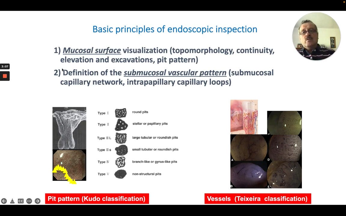
Mucosal Surface Visualization: This is the first essential step and involves a detailed assessment of:
Topomorphology: The overall shape and contours of the mucosal surface.
Continuity: Observing for any breaks, disruptions, or irregularities.
Elevation and Excavations: Identifying any raised areas or depressions.
Pit Pattern: Analyzing the microscopic crypt opening patterns on the mucosal surface, often categorized using the Kudo classification (e.g., Type I: round pits, Type II: stellar/papillary pits, up to Type V: non-structural pits).
Submucosal Vascular Pattern Definition: The second critical component involves evaluating the vessels beneath the mucosal surface:
Submucosal Capillary Network: Assessing the general network of capillaries.
Intrapapillary Capillary Loops (IPCLs): Observing the fine capillary loops within the papillae. This can be assessed using classifications like the Teixeira classification for vessels.
Significance: These two steps are foundational for a comprehensive endoscopic examination, allowing for the identification of subtle changes indicative of early disease.
