

👆 The clip above is a preview from the EndoCollab library. The full lecture, “Endoscopic Characterization of Small Bowel Villi” by Dr. Klaus Mönkemüller, is available to paid EndoCollab members.
The small bowel is not only the body’s largest surface area but also its largest immune organ, housing billions of white blood cells and interacting closely with the microbiota. Despite its importance, inspecting its intricate villous structure can be a challenging task. However, mastering this inspection is key to identifying a range of subtle and significant pathologies.
In this guide, based on a lecture by Dr. Klaus Mönkemüller, we will cover the fundamental principles for characterizing high-quality small bowel villi.
“Advanced Imaging” is a Foundational Skill
The concept of “advanced imaging” is not new; it has been part of endoscopy from the beginning. Dr. Mönkemüller points to a 1974 paper on the “Dye Scattering Method” as an early example of this principle. This early technique involved applying dyes like indigo carmine or methylene blue to enhance mucosal visualization.
Today, this same principle extends to digital chromoendoscopy, but the fundamental goal remains the same: detailed and enhanced visualization of the villi.
Key Steps for High-Quality Villi Observation
Dr. Mönkemüller outlines a systematic approach, shown on his slide, to ensure you can clearly assess the small bowel mucosa.
1. Prepare the View
Remove debris with water.
Remove bubbles, often using simethicone, to clearly appreciate the mucosal surface.
2. Observe Methodically
Always observe before advancing the scope or performing biopsies. This allows you to capture clear images before potential bleeding or manipulation obscures the view.
Avoid too much insufflation, as this can stimulate motility. The speaker notes he prefers to use CO2.
3. Use Augmentation Techniques
Underwater Endoscopy: This technique provides inherent magnification, augmenting visibility by approximately 1.6 times.
Contrast Agents: Using contrast agents allows for better delineation and characterization of mucosal structures.
Artificial Chromoendoscopy: This includes modern technologies like BLI, NBI, FICE, and i-scan.
Capsule Endoscopy: This modality can also provide “marvelous” views of the small bowel.
What Do Normal Villi Look Like?
Normal villi have different lengths and sizes depending on their location in the intestine. Using modern tools like zoom endoscopy (up to 110x magnification) and NBI, you can visualize the intra-villous capillaries and even see the blood flowing through them.
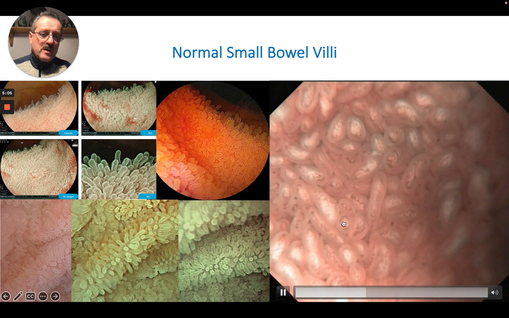
The video clip shows a highly magnified NBI view of normal villi. The villi are distinct, uniform, finger-like projections. Within each individual villus, a dark, looped capillary structure is clearly visible. The speaker narrates that with this view, “the vessels become more visible and clear”.
To see Dr. Mönkemüller’s breakdown of specific pathologic findings in Whipple’s Disease, IPSID, and more, including key visual differentiators, consider becoming a paid subscriber.
