

You’ve just completed a difficult ERCP, sweeping the bile duct multiple times. The final cholangiogram looks clear. But how certain are you that a small, clinically significant stone isn’t hiding in a proximal intrahepatic duct? In many cases, especially after a sphincterotomy, contrast injected at the papilla can flow out too quickly, offering a falsely reassuring image of a clear biliary tree.
There is a simple but powerful technique that can provide definitive answers in these situations and solve several other common diagnostic dilemmas. In this session from EndoCollab, Dr. Klaus Mönkemüller explains that one of the most important and underutilized functions of a standard stone extraction balloon is performing an occlusion cholangiogram.
This article breaks down the mechanics, rationale, and advanced applications of this essential technique, transforming your stone extraction balloon into a powerful diagnostic tool.
The ‘Why’ and ‘How’ of Occlusion Cholangiography
The stone extraction balloon is known for one thing, but it has at least ten distinct uses. The occlusion cholangiogram is arguably one of its most critical secondary functions.
Clinical Rationale: Overcoming Contrast Runoff
The fundamental challenge in many cholangiograms is achieving adequate filling of the biliary tree. After a sphincterotomy, the opening is large, and contrast follows the path of least resistance, flowing out into the duodenum instead of filling the proximal ducts. This can lead to missed stones or incomplete visualization of the anatomy.
The occlusion cholangiogram solves this by using the balloon to temporarily plug the bile duct. This allows for controlled injection of contrast proximally, ensuring the entire biliary system, including the intrahepatic ducts, is fully delineated.
The Technique: Simple Mechanics for a Superior Image
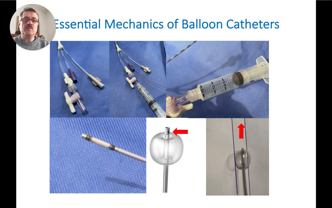
Understanding the mechanics of the balloon catheter is key. Most catheters have two or three ports: one for the guidewire, one to inflate the balloon with air, and sometimes a third dedicated port for contrast injection.
The process is straightforward:
The balloon is advanced to the desired position in the bile duct.
The balloon is inflated via the air port, creating a seal that occludes the duct.
Contrast is then injected. It exits through a specialized opening proximal to the balloon, filling the upstream ducts under controlled pressure.
The primary and most important use for this technique is to confirm the complete clearance of bile duct stones after sweeping.
To see advanced troubleshooting tips, case variations, and a downloadable quick-reference guide based on this video, consider becoming a paid subscriber.
