

You are performing a colonoscopy, and you identify a lesion in the rectum. You snap a photo. But when you look at the image later—or worse, when you write the referral letter for the surgeon—you hesitate.
Is that polyp on the left lateral wall? Or is it anterior?
The flexible nature of the endoscope often disorients our sense of direction, particularly in the large, vault-like space of the anorectum. Yet, accurate localization is critical, especially when guiding future therapy for anal fissures or fistulas.
In this basic endoscopy review, Prof. Klaus Mönkemüller demonstrates a fundamental law of physics that can solve this problem instantly: Water seeks the dependent side.
The Clinical Problem: “Where are we?”
At the start of the procedure, you may encounter multiple polyps.
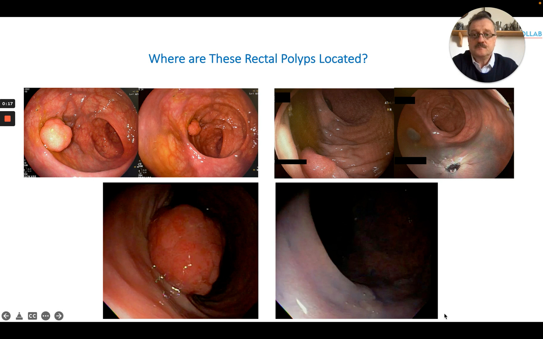
On Screen: Four distinct endoscopic images are displayed simultaneously. They show various rectal polyps of different morphologies (sessile, pedunculated). The camera angle varies in each, making it difficult to intuitively know which wall of the rectum is being viewed based solely on the image.
Prof. Mönkemüller asks the essential question: “Where are these polyps located?”. Without a reference point, it is nearly impossible to tell.
The Technique: The Water Trick
The solution relies on the patient’s position and gravity.
Step 1: Confirm Patient Position Most standard colonoscopies are performed with the patient in the Left Lateral Decubitus position.
Step 2: Instill Water Flush a small amount of water into the rectal vault.
Step 3: Observe the Pooling Because the patient is lying on their left side, gravity will pull the water to the patient’s left. Therefore, wherever the water accumulates on your screen defines the Left side of the patient.
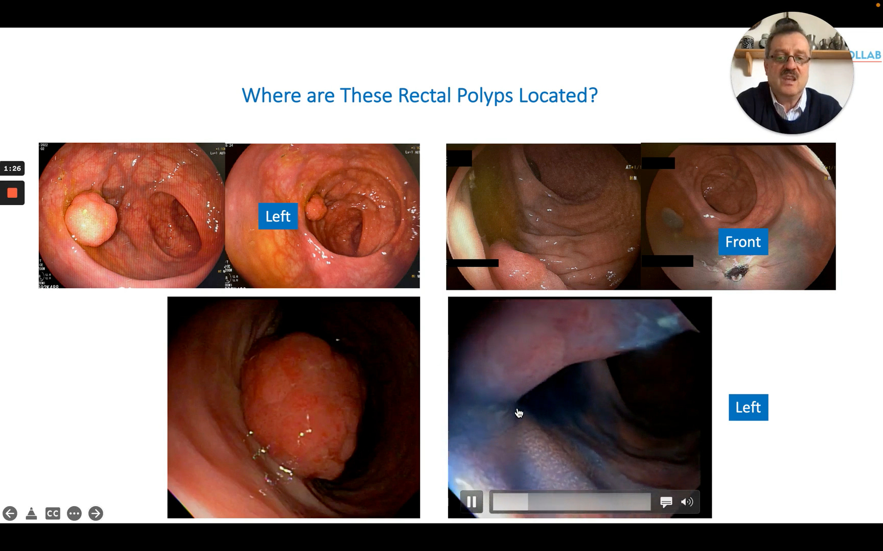
On Screen: A large polyp is visualized. The endoscopist has used chromoendoscopy with blue dye (Indigo Carmine). The blue liquid clearly pools on one side of the lumen. Significance: The dye acts as a high-contrast fluid level. Because the blue fluid pools at the bottom of the gravity well, and the patient is on their left side, the pool of blue dye confirms that wall is the Left wall.
Prof. Mönkemüller notes: “You can see that the water accumulates on the left side. Therefore, the polyp is located on the left.”
Subscribe to EndoCollab to access the complete quadrant mapping guide, including how to identify Anterior vs. Posterior walls using this technique, and to download the Quick Reference Card.
